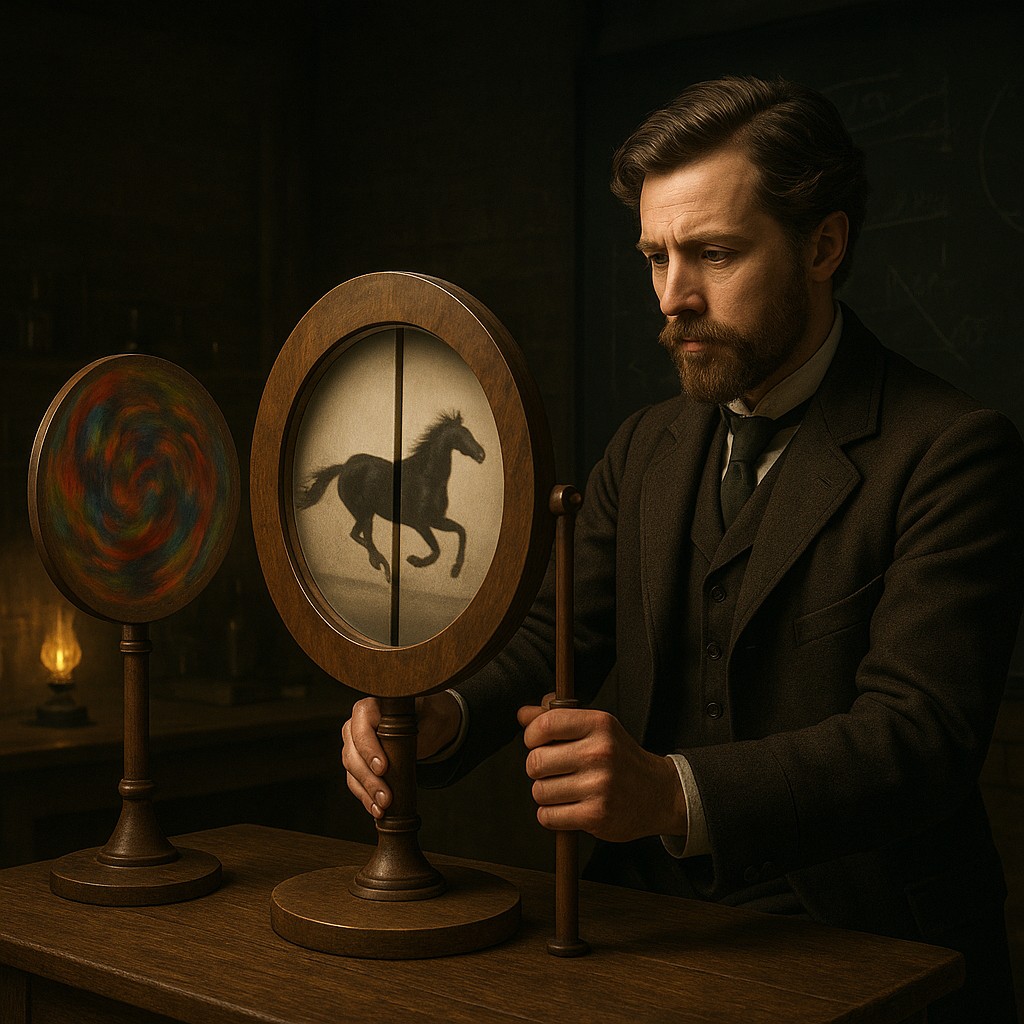
Imagine standing in a dimly lit laboratory in the mid-18th century. You are watching a scientist spin a wooden disc covered in what looks like a smeared, psychedelic mess of ink. The shapes are stretched, bloated, and completely unrecognizable, like something from a distorted funhouse mirror. But then, the scientist places another disc with a single narrow slit in front of the mess and begins to spin it. Suddenly, as if by a digital trick from a century in the future, the chaos vanishes. In its place is a perfectly proportioned, steady image of a galloping horse or a blooming flower. You are witnessing the anorthoscope, a device that proves your eyes do not just see the world - they actively build it.
This phenomenon is more than just an old-fashioned party trick. It represents a fundamental "glitch" in human biology that reveals how we process reality. We often assume our vision works like a video camera, capturing a smooth stream of the world in real time. In truth, our brains are constantly stitching together fragmented snapshots, filling in blanks and ignoring gaps. The anorthoscope exploits a specific biological delay called "persistence of vision." It forces the brain to work overtime to turn a series of disconnected "slices" into a clear picture. By studying this effect, we can pull back the curtain on the internal machinery that allows us to see movement where there is none and stability where there is only a flickering light.
The Architect of the Invisible Image
To understand why a distorted mess can look like a masterpiece, we have to look at the work of Joseph Plateau. He was a Belgian physicist who pioneered this field in the late 1820s. Plateau was obsessed with how the human eye holds onto light. He realized that when light hits the retina (the light-sensitive layer at the back of the eye), the chemical reaction does not stop the instant the light source disappears. Instead, there is a lingering "afterglow" that lasts for a fraction of a second. This is the same reason that if you swirl a sparkler in the backyard at night, you see a glowing circle rather than a single moving dot. Your brain holds onto the previous position of the sparkler while the new one is being recorded.
Plateau’s anorthoscope took this concept to a radical extreme. He created "anamorphic" images, which are drawings intentionally stretched out using precise math. On their own, they look like nonsense. However, when these images spin behind a narrow slit, the eye only sees a tiny vertical sliver of the drawing at any given micro-second. Because the disc moves so fast, the brain receives a rapid-fire sequence of these slivers. Thanks to the persistence of vision, the brain stores the first sliver just long enough to overlap it with the second, third, and fourth. Eventually, it "assembles" the data, and you see the corrected image perfectly centered in the slit.
The Chemistry of the Biological Buffer
The reason this stitching process happens at all is due to how nerve cells communicate with the brain. When particles of light hit the sensors in your eyes, they trigger a chain of electrical signals. This process is incredibly fast, but it is not instant. There is a physiological "fade time." If a second image arrives before the signal from the first image has fully faded, the brain treats them as part of the same event. This is known as "temporal integration." In the context of the anorthoscope, the brain acts as a biological buffer, holding onto bits of data until it has enough information to make sense of the scene.
This is fundamentally different from a digital screen. On a computer monitor, an image stays put because the pixels remain lit. During an anorthoscope viewing, the image is never actually "there" in its entirety at any single moment. You are essentially seeing a ghost - a mental construct built from a dozen different moments in time layered on top of each other. This shows that seeing is not a passive act of receiving light, but an active task of reconstruction. Your brain is less like a mirror and much more like a master film editor, cutting together fragments to create the illusion of a seamless story.
Comparing Visual Perception Technologies
While the anorthoscope might seem like an early version of cinema, the mechanics are different. Understanding these variations helps clarify why the anorthoscope feels so eerie and magical compared to a modern movie or a simple flipbook.
| Device or Effect |
Primary Mechanism |
How It Deceives the Eye |
Result |
| Thaumatrope |
Two-sided spinning card |
Overlapping two distinct images on the retina |
Two images appear to merge (e.g., a bird appearing inside a cage). |
| Phenakistiscope |
Rotating disc with slots |
Rapidly showing slightly different frames |
Creates the illusion of smooth motion from static drawings. |
| Anorthoscope |
Viewing distorted art through a slit |
Combining anamorphic slices over time |
Rebuilds a corrected, stable image from a mangled mess. |
| Modern Cinema |
High-frequency projection |
Uses persistence and the "phi phenomenon" |
Individual frames are perceived as continuous, life-like movement. |
The key lesson here is that while most optical toys focus on creating movement, the anorthoscope focuses on creating form. It is the only device that takes an unrecognizable shape and uses motion to "fix" it. In a way, the motion acts as a filter that decompresses the distorted data. This allows your visual cortex (the part of the brain that processes sight) to reorganize the information into its intended shape.
Beyond the Eye: The Role of the Inferior Temporal Cortex
For a long time, scientists thought this was strictly a trick of the eye - a simple chemical trail left on the retina. However, modern neuroscience tells a more complex story. Recent studies involving brain scans have shown that "slit-viewing" actually uses the inferior temporal cortex. This is the part of the brain responsible for high-level object recognition. It is the part of you that knows a table is a table regardless of the angle you are looking from.
When you look through the slit of an anorthoscope, your inferior temporal cortex goes into overdrive. It receives "partial shape signals." Imagine someone trying to describe a famous painting to you by only showing you one square inch at a time through a hole in a piece of cardboard. If they move the cardboard fast enough, your brain eventually takes a mental shortcut and realizes, "Oh, that’s the Mona Lisa!" This suggests that our ability to recognize objects is not just based on seeing the whole thing at once, but on the brain's ability to predict and combine shapes over time. This "spatiotemporal integration" is the same mechanism that allows you to recognize a car driving past a picket fence, even though the fence blocks 70 percent of the car at any given moment.
The Mathematical Magic of Distortion
The magic of the anorthoscope relies on a specific type of geometry called anamorphic projection. To create an image for the device, an artist cannot simply draw a horse. They must draw a horse that has been mathematically "unwrapped." If the viewing disc and the image disc spin at different speeds, the image must be distorted to match that specific ratio. For example, if the image disc spins twice as fast as the slit disc, the drawing must be compressed or stretched by a factor of two to appear normal to the viewer.
This reveals a fascinating relationship between time and space. In the world of the anorthoscope, time (the speed of the spin) is used to correct space (the distorted drawing). If you change the speed of one disc, the image begins to stretch or shrink before your eyes. This is a physical demonstration of how our perception of an object's size and shape is tied to how fast it is moving relative to us. It challenges the common-sense idea that an object’s properties are fixed. In our minds, what we see is always a calculation involving light, time, and our own biological processing speed.
Common Misconceptions and Optical Myths
One of the most frequent mistakes people make when discussing the anorthoscope is confusing it with a simple blur. We often think that if something moves too fast, it just becomes a smear. But the anorthoscope uses that smear to create clarity. It is not an "optical illusion" in the sense that your eye lens is being tricked by bending light. Instead, it is a "cognitive illusion" where the brain is forced to use its internal logic to solve a puzzle.
Another misconception is that this effect requires high-tech precision. In reality, you can recreate this at home with a piece of cardboard and a fan. The principle is so strong because it is baked into our survival instincts. Our ancestors needed to be able to identify a predator moving through tall grass. Even if they could only see slices of a tiger through the blades, their brains needed to stitch those slices together instantly to recognize the danger. The anorthoscope simply takes that survival mechanism and uses it to play a game with art.
The Future of the Flickering Image
Today, the principles behind the anorthoscope have found their way into technologies that Joseph Plateau could never have imagined. Medical imaging, such as CT scans, works on a similar logic: taking many slices of a human body and using a computer (rather than a brain) to stitch them into a three-dimensional model. Even the way digital video compression works - where only the moving parts of a frame are updated while the rest is held in a "buffer" - echoes the brain's strategy of holding onto visual information to save processing power.
As we move toward a world of augmented reality and high-speed displays, understanding the limits and capabilities of our "biological buffer" is more important than ever. We are learning that the human mind is remarkably flexible, capable of finding order in chaos as long as the timing is right. The anorthoscope serves as a timeless reminder that our window into the world is not a wide-open view, but a narrow slit through which we glimpse fragments of reality. It is our brain's incredible capacity for synthesis that turns those fragments into the beautiful, coherent world we think we see.
Take a moment to consider the sheer processing power happening behind your eyes right now. Every time you blink, every time you turn your head, and every time something zips past your field of vision, your brain performs the same stitching that makes the anorthoscope work. You are not just a spectator of the world; you are its constant, silent architect. Let this realization inspire you to look a little closer at the "blur," because within that chaos, your mind is always busy creating a masterpiece of order. Embrace the flicker, for it is in those tiny gaps of time that the true magic of human consciousness resides.