Why understanding how the eye turns light into images changes everything
Looking is something we do thousands of times a day without thinking. Yet that simple act is the result of a lightning-fast, highly organized chain of events, from capturing photons in the environment to your brain assembling an image. Learning how the eye works is not just scientific curiosity, it helps you understand perception, eye health, and even fields like photography and artificial intelligence. In short, biology explains the everyday magic.
Imagine a dark room that lights up the instant you flip a switch. The eye does that, and much more: it controls how much light enters, focuses fine detail, detects color, and encodes shapes so your brain can interpret a scene. Each step follows its own rules and trade-offs, adding either useful information or noise to the final image. Understanding these steps explains why certain visual problems occur, why night changes what you see, and why two people can literally see the same scene differently.
This text will walk you through the process step by step. We start with eye anatomy, then cover the optical principles that form an image on the retina, move on to how photons become electrical signals, and finish with how the brain assembles those signals into a coherent perception. The style is clear, full of concrete examples, and a little playful when it helps memory. By the end, you will have practical ways to explain vision to someone else, and you will be able to correct some common misconceptions.
Get ready to discover that the eye is not a perfect camera, that "resolution" varies with the distribution of cells, and that what you "see" is as much constructed by your brain as captured by your eye. It is mechanical, chemical, and deeply computational. Let’s go.
The structure of the eye explained without complicated jargon
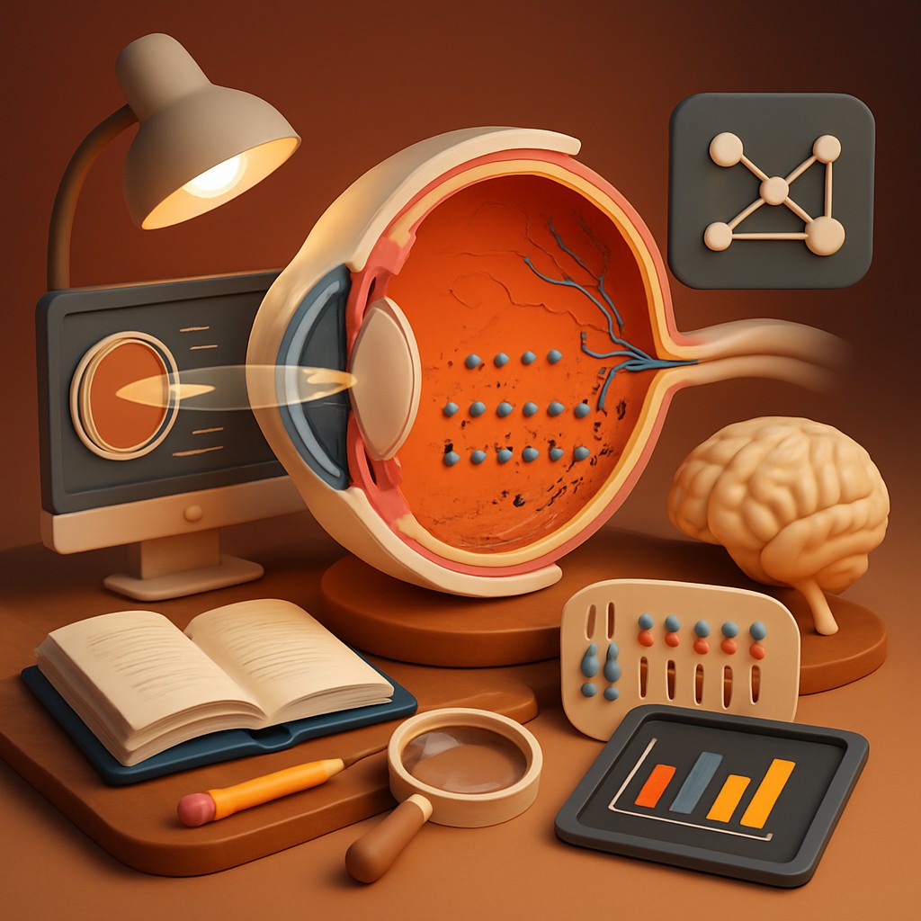
From the outside, the eye looks like a fluid-filled sphere with a front window that lets light in. That window is the cornea, a transparent surface that bends incoming light. Behind the cornea is the iris - the colored part - which adjusts the opening called the pupil, a bit like a camera lens controlling light levels.
Behind the iris sits the lens, a small flexible structure that changes shape to focus on near or far objects. Together, cornea and lens focus light onto the retina, the photosensitive tissue lining the back of the eye. The retina is the real star: it is where light is converted into electrical signals.
Those signals then gather and leave the eye via the optic nerve, bound for the brain. A few accessory structures are worth mentioning: the aqueous humor and the vitreous body, which maintain the eye’s shape, and the conjunctiva, which protects the surface. Each part has a precise role, and a small change in any of them can alter what you see.
How light becomes a sharp image: simple optical principles
When light enters the eye, it carries an inverted image of the outside scene because of the curvature of the optical surfaces. The cornea and lens bend the light rays so they converge on the retina. If convergence is perfect, the focal image lands exactly on the layer with photoreceptors, and you get sharp vision.
The lens changes thickness thanks to muscles called the ciliary muscles to focus on close objects - this is accommodation. With age, the lens loses flexibility and becomes less able to thicken, which explains presbyopia, the difficulty reading up close after age 40-50. Focusing errors like myopia and hyperopia result from an eye that is too long or too short respectively, or from an improperly curved cornea. Glasses or contact lenses correct these errors by altering the path of light rays.
The iris acts like an aperture: it narrows the pupil in bright light to protect the retina and increase depth of field, and it widens in low light to let in more photons. But enlarging the pupil also increases optical aberration, which can make the image less sharp. The visual system balances these trade-offs automatically.
Photoreceptors: the chemical heart of vision
The retina contains two major families of light-sensitive cells: rods and cones. Rods are ultra-sensitive and excellent for low-light vision, but they do not discriminate color well. Cones, by contrast, are less light-sensitive but responsible for color vision and fine detail. The fovea, a small central region of the retina, is densely packed with cones and provides the highest visual acuity.
Each photoreceptor contains visual pigments that change shape when a photon is absorbed, triggering a chemical cascade called phototransduction. This cascade converts light information into a modulated electrical signal. The signals from millions of photoreceptors are then processed by layers of retinal neurons - bipolar cells, ganglion cells, and horizontal cells - which perform early image processing like contrast detection and redundancy reduction.
The photon-to-signal conversion is remarkably fast and precise. However, it is not perfect: there is always some noise (for example thermal noise in the pigments) and statistical limits set by the number of available photons, especially in low light. The system compensates by integrating signals over time and space to smooth the output, but these compensations influence the final perception.
From retinal signal to brain: the path and sorting of information
The axons of the ganglion cells bundle together to form the optic nerve. Fibers in the optic nerve leave the eye and partially cross at the optic chiasm, allowing a geographic distribution of visual information to the brain hemispheres. Thus, the right half of the visual field is processed mainly by the left hemisphere, and vice versa.
After the chiasm, signals travel through the lateral geniculate nucleus - a relay station in the thalamus - before reaching the primary visual cortex in the occipital lobe. The visual cortex does more than display the image; it breaks it down into elements - orientation, motion, depth, color - across several specialized areas. These pieces are then recombined and integrated with memory, attention, and other senses to produce conscious perception.
This process is highly parallel and hierarchical. Early layers extract simple features, and higher layers build objects and scenes. There are also feedback loops: the brain directs attention to certain elements and modulates activity in the retina and visual areas based on perceived importance.
Perception, illusions, and what the brain "fills in"
What you see is not a perfect photograph of the world, but an interpretation. The brain uses probabilistic shortcuts, called heuristics, to infer reality from partial or ambiguous information. These heuristics sometimes produce fascinating illusions, like straight lines that appear curved, colors that shift depending on context, or objects that vanish if you do not look directly at them.
A common misconception is that the eye takes complete images and sends them "as is" to the brain. In reality, most fine detail is perceived only where the eyes are directed, and the brain "fills in" peripheral areas from cues and internal models. Perception also depends on context: the same gray can look different depending on surrounding colors.
Another surprising point is that processing speed is not uniform: the system is incredibly fast at detecting motion and direction, but slower at judging complex details and spatial relations. This explains why we react almost instantly to a ball thrown at our face, yet take longer to decipher an unreadable text.
Useful summary table: rods vs cones and primary roles
| Characteristic |
Rods |
Cones |
| Light sensitivity |
Very high, excellent in low light |
Less sensitive, better in bright light |
| Color perception |
No, grayscale vision |
Yes, trichromatic (red, green, blue) |
| Predominant location |
Retinal periphery |
Fovea and central regions |
| Spatial resolution |
Low, not for fine detail |
High, enables fine acuity |
| Main function |
Motion detection and night vision |
Distinguish colors and details |
| Approximate number |
~120 million |
~6 million |
This table explains why your night vision is blurry and colorless, while reading and face recognition rely on the cone-rich central retina.
Practical applications and memory tricks
Knowing how the eye works has practical uses: choosing glasses, understanding why a bright screen tires you, or optimizing lighting for work. For example, eye strain often comes from poor contrast and excessive exposure to high-frequency light - adjusting brightness, taking breaks, and blinking more frequently really helps.
To remember the steps, use a small mental story: light enters through the "door" cornea, passes the "curtain" iris, goes through the "zoom" lens, is imprinted on the "film" retina, then the "messenger" optic nerve carries the information to the "studio" cortex for final editing. This simple image helps recall the physico-chemical sequence and the role of each part.
Quick eye-health tips: take regular breaks when using screens (20-20-20 rule: every 20 minutes, look at something 20 feet away for 20 seconds), protect your eyes from the sun with UV sunglasses, and see a specialist if your vision changes suddenly. Many visual problems are easier to fix when detected early.
Answers to common misconceptions
Misconception 1 - "We only use 10% of our vision." False. You use all of your vision, but different regions of the retina specialize in different tasks. The illusion comes from the fovea capturing most fine detail, making the rest seem "less important."
Misconception 2 - "Glasses make your eyes lazy." Partly false. Glasses do not make the eyes weaker by themselves, but they change how the eye muscles work. In some cases, correcting a strong difference between the two eyes can restore normal coordination.
Misconception 3 - "Blinking is just for moistening the eye." Blinking mainly lubricates, but it also helps reset visual attention and reduce glare. A blink is a tiny, useful reset.
Small experiments to try at home to understand better
There are simple and safe experiments to feel how the eyes and brain cooperate. For example, stare at a central cross and notice how peripheral bright spots seem to disappear - that is an effect of central focus. Another experiment is to switch focus between a nearby and a distant object to feel accommodation effort. Finally, observe how color perception changes under different lighting to understand chromatic adaptation.
These small exercises help anchor abstract concepts in sensory experience. They also show perception variability: different people may report slightly different impressions, reminding you that seeing is both physiological and interpretive.
In conclusion: why this knowledge makes you smarter and more curious
Understanding how the eye turns light into images gives you a fresh perspective on the everyday. You will see light as data, and vision as a series of graceful biological computations. This knowledge lets you explain why glasses help, why night erases color, and why an illusion can fool you in milliseconds.
More importantly, it sparks curiosity: who would have thought that reading is a cultural technology that exploits the fovea and your brain's ability to recognize patterns? Learning about vision gives you practical tools to protect your eyes, optimize your visual environment, and appreciate the biological engineering that lets you admire a sunset.
Go ahead, look around with a new eye - literally and figuratively. You will better understand visual impressions, correct misconceptions, and be able to share these discoveries with others, which is the best way to learn.